


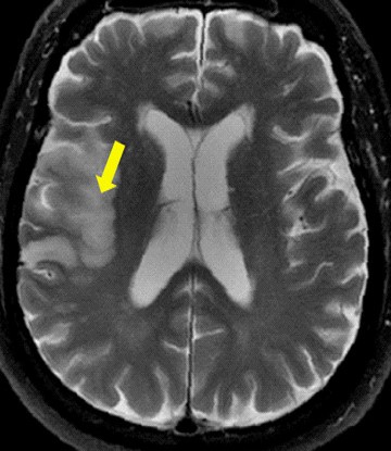
Magnetic resonance imaging showing cerebral infarction.
Severe stenosis of major intracranial arteries increases the risk of cerebral infarction after revascularization surgery in moyamoya disease
CHINA, July 20, 2026 /EINPresswire.com/ -- Currently, the treatment for moyamoya disease (MMD), a condition characterized by the blockade of the internal carotid artery, is revascularization surgery. However, this surgery can lead to cerebral ischemia for which limited predictive tools are available. Now, a group of Chinese researchers identified preoperative blockage of the major intracranial arteries as a risk factor for cerebral infarction after combined revascularization surgery in MMD, paving the way for the development of novel prediction protocols.The buildup of fats, cholesterol, calcium, and immune cells within the blood vessel can lead to the narrowing or even complete blockage of the blood vessel. This condition, called stenosis, can lead to various disorders. Moyamoya disease (MMD) results from stenosis of the blood vessel in the neck that supplies blood to the brain (distal internal carotid artery (ICA)), as well as its branches. This leads to the suboptimal blood supply to the brain, a condition termed cerebral ischemia. Currently, the main treatment strategy for MMD involves the surgical augmentation of blood supply to the brain (neovascularization). However, neovascularization is associated with cerebral ischemia risks. The prediction of cerebral ischemic events after surgery has been challenging, mainly because the related risk factors have not been identified.
To address this knowledge gap, a research team led by Dr. Yuanli Zhao and Prof. Rong Wang examined the medical imaging features of 119 patients with ischemic MMD who underwent neovascularization surgery at the Department of Neurosurgery, Beijing Tiantan Hospital, Capital Medical University. The study was published in the Chinese Neurosurgical Journal on 14 April 2026. Commenting on the rationale of the study, Dr. Zhao remarked, “The incidence of cerebral ischemia after revascularization surgery in MMD is about 5%. The identification of risk factors for postoperative cerebral ischemia can aid in developing predictive tools and consequently improving patient outcomes.”
All patients in this study underwent digital subtraction angiography (DSA), magnetic resonance imaging (MRI), and computerized tomography (CT) perfusion before surgery. In DSA, the blood vessel of the brain is visualized using dye injected via a catheter. After the neovascularization surgery, the patients underwent CT at 4–6 h post-surgery and head MRI at days 5–7 post-surgery. Cerebral artery blockage was quantified using the Suzuki staging system. Oxygen deprivation-induced tissue death (infarction) was identified based on a low-density signal in the CT scan or a prominent signal in the MRI. The researchers examined postoperative infarctions around major arteries. New infarctions developed in 11 hemispheres in 8 patients (5.5% of the patients), while infarctions on both sides of the brain were observed in 3 patients. Severe blockage of the major cerebral arteries (anterior, middle, and posterior cerebral arteries) was associated with postoperative infarction.
DSA of cerebral arteries was performed for 3 patients who presented with cerebral ischemia after the surgery. In these patients, brain imaging before the surgery was characterized by severe occlusion of the major arteries. This confirmed the preoperative occlusion of the major arteries as a risk factor for postoperative cerebral infarction. According to Prof. Wang, “The findings of this study can aid clinicians to predict cerebral ischemia events after revascularization surgery and implement relevant mitigation strategies, improving the treatment outcomes.”
Although this study identified a risk factor for postoperative cerebral infarction, the roles of other confounding factors, such as age and surgery side, cannot be ruled out, warranting further studies with a larger sample size. However, this study has laid the foundation for evaluating other risk factors and developing therapeutic or management strategies to minimize the incidence of postoperative cerebral infarction in patients with MMD.
***
Reference
Title of original paper: Severe stenosis of major intracranial arteries: an important risk factor for infarction complications after combined revascularization in adult patients with ischemic moyamoya disease
Journal: Chinese Neurosurgical Journal
DOI: 10.1186/s41016-026-00430-0
About Capital Medical University
Capital Medical University, Beijing, China, was founded in 1960. The main campus of the school has 12 colleges and 2 research centers. The school also has 41 clinical colleges and departments, and 40 clinical diagnosis, treatment and research centers. The school currently has 16,763 full-time students, including 8,548 graduate students, 7,559 undergraduate students, 261 vocational college students, 395 international students, and 2,605 adult education students. The school and its affiliated hospitals currently have 9,158 teachers, including 9 academicians; 1,296 professors and 2,294 associate professors; 1,554 doctoral supervisors and 2,039 master's supervisors.
Website: https://www.ccmu.edu.cn/
About Prof. Rong Wang from Capital Medical University
Dr. Rong Wang is a professor and neurosurgeon affiliated with the Department of Neurosurgery at Capital Medical University and Beijing Tiantan Hospital. His research interests include cerebrovascular diseases, especially moyamoya disease, intracranial aneurysms, cerebral revascularization, and neurovascular surgery. He is widely recognized for his contributions to understanding the pathophysiology, imaging, surgical management, and outcomes of complex cerebrovascular disorders. His work integrates clinical research with advanced neuroimaging and translational approaches to improve diagnosis, risk stratification, and treatment strategies for patients with cerebrovascular disease.
Yi Lu
Chinese Neurosurgical Journal
+861059978478 ext.
luyi617@sina.cn
Legal Disclaimer:
EIN Presswire provides this news content "as is" without warranty of any kind. We do not accept any responsibility or liability for the accuracy, content, images, videos, licenses, completeness, legality, or reliability of the information contained in this article. If you have any complaints or copyright issues related to this article, kindly contact the author above.
